 |
SECTIONING We have developed a technique of sectioning tissue pieces that allows visualizing in SEM structures lying
underneath the surface, such as epithelial basement membrane, stromal cells, vessels and other compartments, or even
inside the cells. In most cases, a section runs through the epithelial cells exposing intracellular structures:
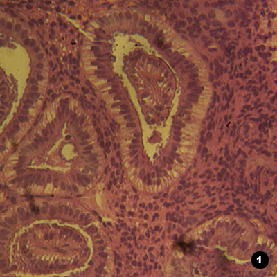
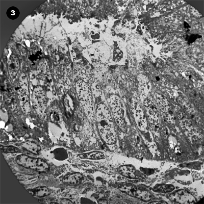
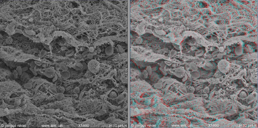
Endometrial gland surrounded by its basement membrane and stroma, exposed by the sectioning technique. This endometrium was
infected and the lumen filled with mucus containing particulate matter.
|
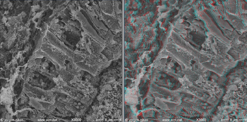
Detail of the above figure. Epithelial cells are cut exposing intracellular structures and organelles. The basement membrane is on the left, and the apical membrane on the right, towards the lumen.
|
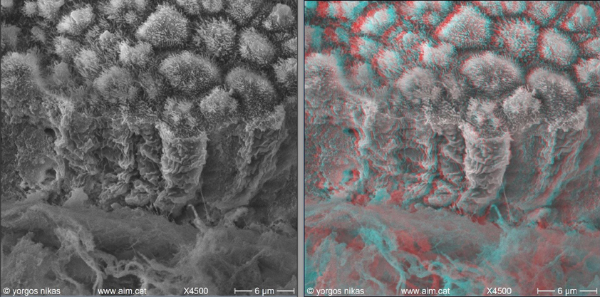
Rarely, a section exposes the lateral epithelial cell membranes intact. A basement membrane sheath lies at bottom.
|
|
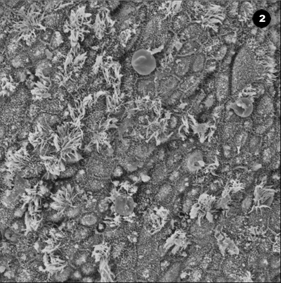
Endometrial stroma exposed by sectioning. Some narrow corridors are seen containing red and white blood cells.
|
|
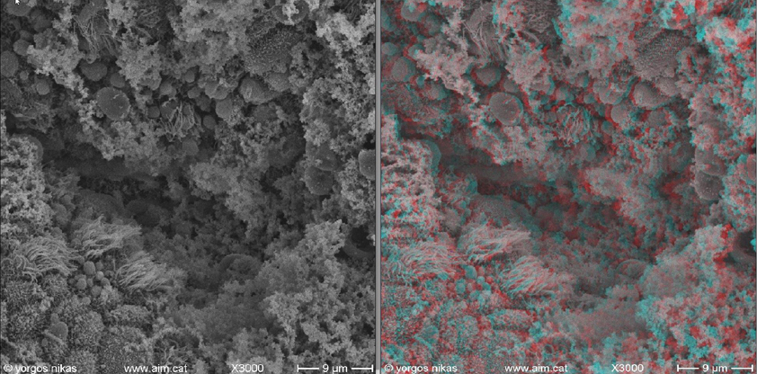
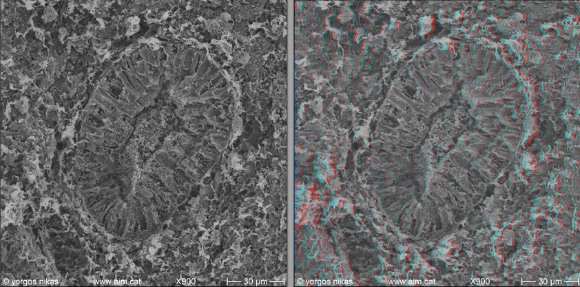
When two "sister" sectioned pieces are viewed stereoscopically side by side, it becomes obvious that their surfaces are
perfectly complimentary and unspoiled from the cut. |
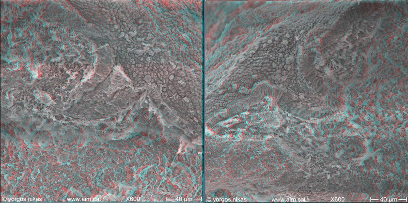
Two "sister" pieces of endometrium obtained by sectioning. The cut runs through an endometrial gland. |
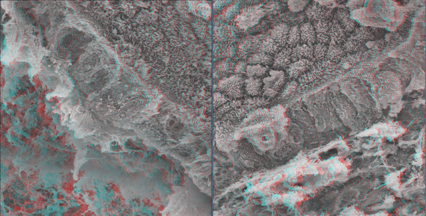
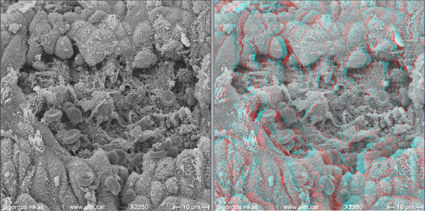
Detail of the above figure, displaying the sectioned epithelium. Except for some slight displacement of the basement membrane, the newly exposed surfaces show absolute complementarity.
|